Wellcome uses cookies.
Read our policy
Close cookie notification
Skip to main content
Wellcome Collection homepage
Visit us
What’s on
Stories
Collections
Get involved
About us
Sign in to your library account
Search our stories, images, catalogue and events
Library account
Search our stories, images, catalogue and events
Search
Images search
Search for images
Search
All
Stories
Images
Catalogue
Events
Colours
Licences
Creative Commons CC-BY (97)
Public Domain Mark (64)
Creative Commons CC-BY-NC (24)
Creative Commons CC0 (14)
In copyright (8)
Creative Commons CC-BY-NC-ND (1)
Dates
From
to
Types/Techniques
Engravings (14)
Ephemera (7)
Leaflets (4)
Lithographs (4)
Paintings (4)
Periodicals (3)
Photomechanical prints (3)
Posters (3)
Prints (3)
Advertisements (2)
Cigarette cards (2)
Drawings (2)
Ink drawings (2)
Oil paintings (2)
Portrait paintings (2)
Watercolors (2)
Allegorical prints (1)
Broadsides (1)
Mezzotints (1)
Periodical (1)
Subjects
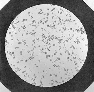
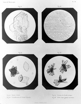
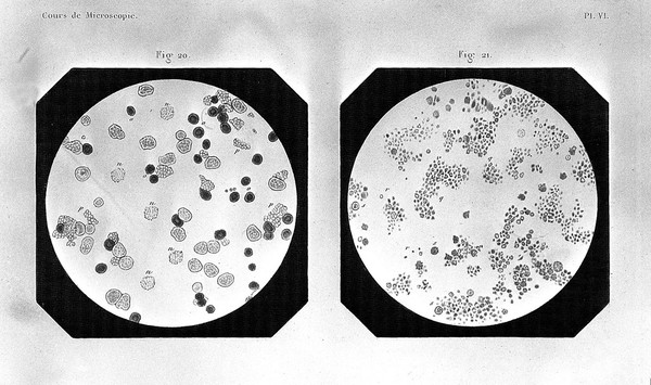
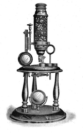
Microscopy (75)
Natural History (33)
Blue (25)
Red (23)
Green (17)
Clusters (14)
Cancer (12)
Network (12)
Circles (10)
Connections (10)
Immune response (10)
Protection (10)
Purple (10)
Barriers (9)
Immunology (9)
Leucocyte (9)
Mouse (9)
Physiology (9)
Structure (9)
T cell (9)
Contributors
Fernan Federici & Jim Haseloff (30)
Hooke, Robert, 1635-1703 (22)
Wellcome Images (14)
Dr. Xiao-nong Wang, Human Dendritic Cell Laboratory, Newcastle University (9)
Leeuwenhoek, Antoni van, 1632-1723 (9)
William R. Geddie (9)
Baker, Henry, 1698-1774 (8)
Blankaart, Steven, 1650-1702 (6)
Bell, Andrew, 1726-1809 (5)
Bidloo, Govard, 1649-1713 (5)
Donné, Alfred, 1801-1878 (5)
Foucault, Léon, 1819-1868 (5)
Marton, Claire K (5)
Marton, L. (Ladislaus), 1901- (5)
Quekett, John, 1815-1861 (5)
Samantha Krukowski (5)
Addey, Frederick (4)
Lecture League (4)
NIMR, MRC (4)
Khuloud T. Al-Jamal & Houmam Kafa (3)
Submit
235 results
Search result sorting
Sort by:
Relevance
Production dates
Sort order:
Ascending
Descending
Submit
Previous (page 1)
Page
2
of 8
Next (page 3)
Close modal window
Previous (page 1)
Page
2
of 8
Next (page 3)


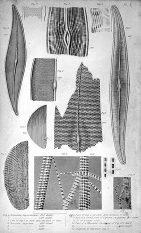


![Martin Frobenius Ledermüllers ... [Vol. I.] Mikroskopische Gemüths- und Augen-Ergötzung: bestehend in ein hundert nach der Natur gezeichneten und mit farben erleuchteten Kupfertafeln, sammt deren Erklärung. [Vol. II] Nachlesse, etc. I[-V] Sammlung.--Abgenöthigte Vertheidigung; als ein Anhang seiner Mikroskopischen Gemüths- und Augen-Ergötzung: wider einige von dem ... Verfasser des Neuesten aus dem Reiche der Pflanzen [i.e. W.F. von Gleichen] ... geäuserte Zweifel und Vorwürfe. Nebst einer Beylage ... A.W. Winterschmidts, welche die Abbildung einer ... Stubenfliege, enthält / [Martin Frobenius Ledermüller].](https://iiif.wellcomecollection.org/image/L0002486/full/282%2C/0/default.jpg)
